
ABI, Ultrasound Color Doppler, Angiography
- Home
- Vascular Treatment
- ABI, Ultrasound Color Doppler, Angiography
Here’s what we can gather about ABI, Ultrasound Color Doppler, and Angiography primarily based on the search outcomes:
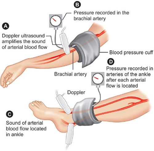
ABI (Ankle-Brachial Index): ABI is a non-invasive test that compares the blood strain in the ankle with the blood strain within the arm. It is used to assess the blood glide and stumble on peripheral arterial ailment (PAD) within the decreased extremities. A lower ABI value suggests decreased blood glide and capacity blockages within the arteries of the legs or feet. ABI is typically performed the usage of ultrasound and is useful in evaluating the chance of PAD, heart attack, stroke, and limb issues.
Ultrasound Color Doppler: Ultrasound Color Doppler is a diagnostic imaging method that mixes conventional ultrasound imaging with shade mapping to visualize blood drift in the body. It uses sound waves to create real-time images of blood vessels and organs, whilst shade mapping represents the direction and speed of blood going with the flow. Colour Doppler is normally used to evaluate blood glide in various clinical conditions, including peripheral arterial disease, deep vein thrombosis, and arterial stenosis.
Angiography: Angiography is an imaging approach that involves the injection of a comparison dye into blood vessels to visualize their structure and blood glide. It is generally used to diagnose and evaluate situations together with arterial blockages, aneurysms, and vascular malformations. Angiography can be executed using special methods, along with traditional X-ray angiography, computed tomography angiography (CTA), and magnetic resonance angiography (MRA).
Understanding ABI, Ultrasound Color Doppler, Angiography
Based on the results, here’s a summary of the statistics about ABI, Ultrasound Color Doppler, and Angiography:
ABI (Ankle-Brachial Index): ABI is a non-invasive check that compares the blood pressure within the ankle with the blood stress within the arm. It is used to assess the blood that goes with the flow and stumble on peripheral arterial disease (PAD) inside the decreased extremities. ABI is typically finished with the usage of ultrasound and is helpful in evaluating the danger of PAD, coronary heart assault, stroke, and limb issues.
Ultrasound Color Doppler: Ultrasound Color Doppler is a diagnostic imaging method that mixes conventional ultrasound imaging with colour mapping to visualise blood flow in the frame. It is regularly used to assess heart and valve characteristics, assess vascular entry to in sufferers on dialysis, and diagnose situations which include peripheral arterial ailment and arterial stenosis .
Angiography: Angiography is an imaging method that involves the injection of a contrast dye into blood vessels to visualise their structure and blood waft. It is typically used to diagnose and compare situations consisting of arterial blockages, aneurysms, and vascular malformations. Angiography can be finished with the usage of special strategies, consisting of traditional X-ray angiography, computed tomography angiography (CTA), and magnetic resonance angiography (MRA).
These techniques are used by healthcare specialists, which include vascular surgeons and radiologists, to evaluate and diagnose diverse vascular conditions. They offer valuable data approximately blood drift, vessel structure, and capacity abnormalities.
-
What is color Doppler ultrasound used for?
In addition to finding clots, Doppler ultrasound can be used to: Check blood flow in your veins, arteries, and heart. Look for narrowed or blocked arteries. See how blood flows after treatment.
-
What is the meaning of angiogram and Doppler ultrasound?
An angiography involves injecting dye into the blood vessels so that they show up clearly on X-ray images. A Doppler ultrasound test also may help check for injuries to your arteries or to monitor certain treatments to your veins and arteries.
-
Can color Doppler detect heart blockage?
An Angiography is the only test that confirms blocks in the heart. 2D Echocardiography and color doppler is the only test that can help in making a diagnosis of heart failure because heart failure diagnosis will be done depending on heart function.
-
How to check heart blockage without angiography?
Echocardiography utilizes sound waves to create images of the heart, allowing visualization of its structures and function. Doppler echocardiography assesses blood flow within the heart chambers and can detect abnormalities such as wall motion abnormalities or valve dysfunction associated with CAD.
-
What is the difference between an arterial Doppler and an ABI?
Arterial duplex ultrasound allows you to view the arterial walls and lumen. It also records waveforms and velocities of blood flow to determine the level and extent of arterial disease. In contrast, an ABI test only gives you information about a circulation problem and the degree of disease—but not the location.